Nobel Prize in Chemistry 2004. The Mechanism of ATP-Dependent Degradation of Proteins Involving Ubiquitin
Ya. E. Dunaevsky* and M. A. Belozersky
Belozersky Institute of Physico-Chemical Biology, Lomonosov Moscow State University, 119992 Moscow, Russia; fax: (7-095) 939-3181; E-mail: dun@belozersky.msu.ru* To whom correspondence should be addressed.
Received November 1, 2004
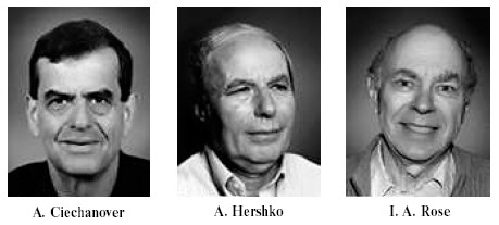
The decision of the Nobel Prize committee in awarding the Nobel Prize in Chemistry for 2004 attracted much attention of journalists. Headlines “kiss of death”, “the black label”, and “death label” clearly and literally underline the scientific problem that has been recognized by this award. As indicated in a press release of the Swedish Royal Academy of Sciences, the winners of the 2004 prize have provided scientific background for understanding the molecular basis of the mechanism of selective degradation of proteins. Two Israeli scientists Aaron Ciechanover and Avram Hershko from Technion Institute of Technology (Haifa) and American scientist Irvin Rose from University of California (Irvine, CA) discovered one of the most important processes occurring in eukaryotic cells, ATP-dependent ubiquitin-mediated protein cleavage [1-10]. This process consists of two steps. The first step includes elucidation and labeling of proteins, which should be degraded. The second step represents degradation of the targeted proteins catalyzed by a large cytosolic protein complex named the proteasome by Goldberg (another distinguished expert in this field) and his colleagues [11]. The ubiquitin-proteasome system provides “coexistence” between intracellular proteins and a non-lysosomal proteolytic system, which can potentially degrade any protein in the cell.
At the current stage of our knowledge, there is the following outline of the process of ubiquitin-mediated proteolysis. Nature developed a highly specialized ubiquitin-conjugating system for identification and labeling of proteins that should be degraded by cells. Special degradation signals recognized by this system may include primary structure motifs (N-terminal amino acid residues), secondary post-translational protein modifications (e.g., phosphorylation), and association with some additional proteins such as viral oncoproteins and molecular chaperones. The protein conjugating system is functionally and spatially proteasome-independent. Proteins identified by the cell as the targets for proteolysis are labeled by covalent attachment of ubiquitin, a small and highly stable protein that consists of 76 amino acid residues. Ubiquitin conjugation (also known as ubiquitination) with protein substrates involves a chain of reactions. Initially ubiquitin activating enzyme (E1) hydrolyzes ATP and forms a thioester bond with ubiquitin. The activated ubiquitin is then transported to one of several ubiquitin-conjugating enzymes (E2) responsible for ubiquitin transport to a substrate specifically recognized by one of the ubiquitin ligases (E3). The last stage of ubiquitination process is characterized by several variants; they depend on E3 membership in a certain family--ubiquitin can be transferred from E2 to E3 before its conjugation with substrate, or activated ubiquitin is directly transported from E2 to E3-bound substrate. It should be noted that E2 and E3 exist as large protein families and members of each family differ in their properties and intracellular localization. They can be involved in various cellular processes such as DNA repair, peroxisome biogenesis, resistance to heat shock, cadmium, etc. It is suggested that various combinations of E2 with various E3 forms playing the key role in specific substrate recognition determine high specificity of ubiquitination process. Ubiquitin bound to protein substrates acts (at least partially) as a sorting signal driving conjugates to a proteasome. Ubiquitin molecules are attached to protein substrate as a polyubiquitin chain (a minimal signal includes four ubiquitin molecules). The latter represent the degradation signal recognized by the proteasome.
Subsequently, proteins labeled by the polyubiquitin chains are cleaved by the proteasome in an ATP-dependent reaction, which terminates by formation of small peptides. At the first moment, ubiquitin recognized by one proteasome subunit is anchored on it until protein unfolding and translocation to a central cavity of the proteasome occur. During protein degradation, polyubiquitin chains being bound to receptor are released from the substrate and cleaved into monomers by isopeptidases. These enzymes are obviously required for receptor clearance from polyubiquitin and recruitment of ubiquitin molecules for subsequent use in the ubiquitination process. The substrate released from ubiquitin is subjected to severe proteolytic degradation by the proteasome complex exhibiting wide substrate specificity.
Studies by the laureates for 2004 clarified the role of protein ubiquitination accompanied by its subsequent degradation in many important processes in the cell [12-14]. These include regulation of transcription factor activity, regulation of cell cycle (transition from G1 to S-phase), degradation of oncoproteins and tumor suppressor proteins, and immune system functioning. There is increasing evidence that genetic changes in protein substrates and/or enzymes responsible for ubiquitin conjugation are associated with Alzheimer disease, Angelman syndrome (a mental retardation), cystic fibrosis, etc.
REFERENCES
1.Ciechanover, A., Hod, Y., and Hershko, A. (1978)
Biochem. Biophys. Res. Commun., 81, 1100-1105.
2.Hershko, A., Ciechanover, A., and Rose, I. A.
(1979) Proc. Natl. Acad. Sci. USA, 76, 3107-3110.
3.Hershko, A., Ciechanover, A., Heller, H., Haas, A.
L., and Rose, I. A. (1980) Proc. Natl. Acad. Sci. USA,
77, 1783-1786.
4.Haas, A. L., Warms, J. V. B., Hershko, A., and
Rose, I. A. (1982) J. Biol. Chem., 257, 2543-2548.
5.Hershko, A., and Ciechanover, A. (1982) Annu.
Rev. Biochem., 51, 335-364.
6.Hershko, A. (1983) Cell, 34,
11-12.
7.Rose, I. A., and Warms, J. V. B. (1983)
Biochemistry, 22, 4234-4237.
8.Eytan, E., Ganoth, D., Armon, T., and Hershko, A.
(1989) Proc. Natl. Acad. Sci. USA, 86, 7751-7755.
9.Hadari, T., Warms, J. V. B., Rose, I. A., and
Hershko, A. (1992) J. Biol. Chem., 267, 719-727.
10.Eytan, E., Armon, T., Heller, H., Beck, S., and
Hershko, A. (1993) J. Biol. Chem., 268, 4668-4674.
11.Arrigo, A. P., Tanaka, K., Goldberg, A. L., and
Welch, W. J. (1988) Nature, 331, 192-194.
12.Hershko, A., Ganoth, D., Pehrson, J., Palazzo, R.
E., and Cohen, L. H. (1991) J. Biol. Chem., 266,
16376-16379.
13.Ciechanover, A., DiGiuseppe, J. A., Bercovich,
B., Orian, A., Richter, J. D., Schwartz, A. L., and Brodeur, G. M.
(1991) Proc. Natl. Acad. Sci. USA, 88, 139-143.
14.Ciechanover, A., Shkedy, D., Oren, M., and
Bercovich, B. (1994) J. Biol. Chem., 269, 9582-9589.